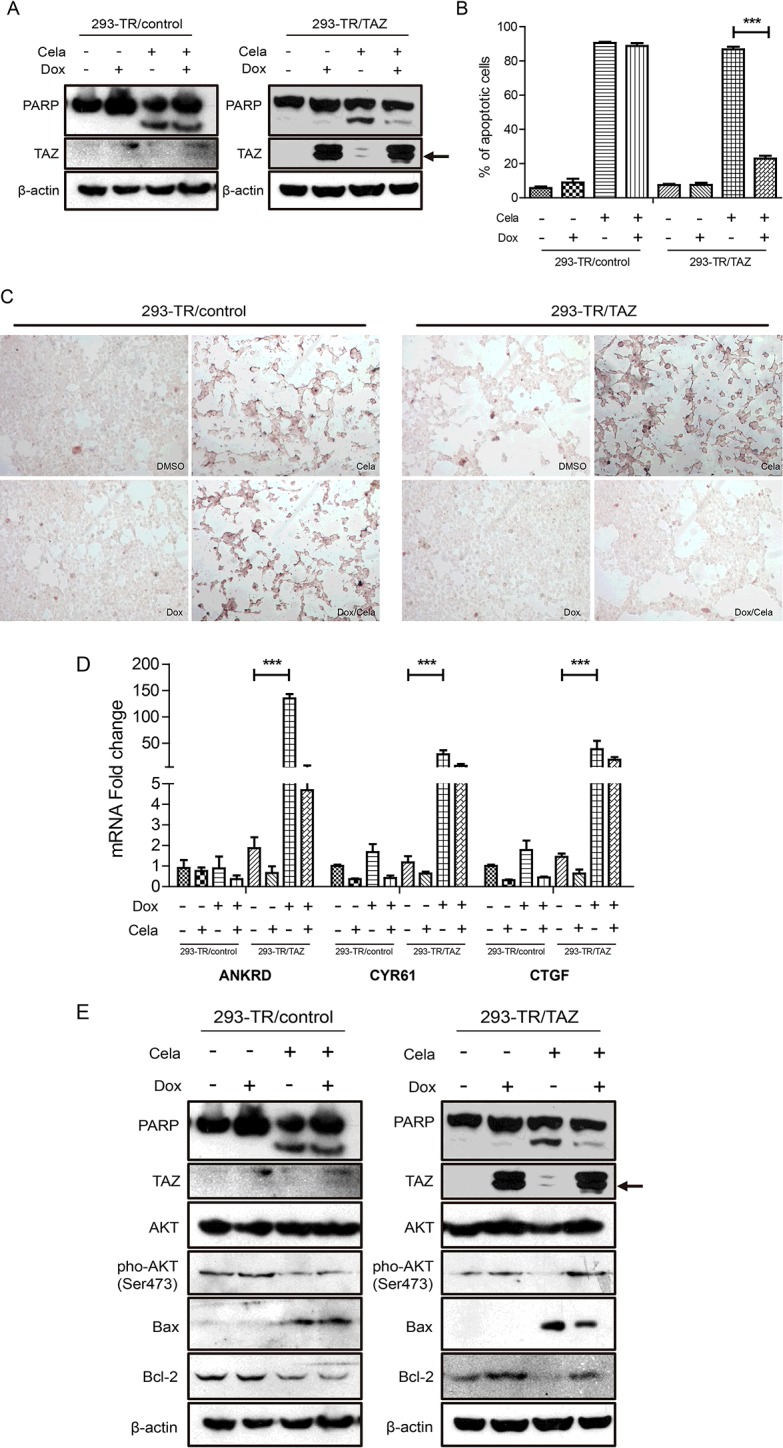
Figure 3. Overexpression of TAZ could attenuate Celastrol-induced apoptosis in T-Rex-293 cells.

(A) Celastrol (0.5 μM) and DMSO were added to 293-TR/control and 293-TR/TAZ cells in the absence and presence of Dox for 48 h. TAZ and cleaved PARP were detected by western blot. β-Actin was used as a loading control. (B) Celastrol (0.5 μM) and DMSO were added to 293-TR/control and 293-TR/TAZ cells in the absence and presence of Dox for 48 h respectively. TUNEL assay was used to detect the apoptotic phenotype. TUNEL-positive (apoptotic) cells were stained brown (magnification 100 ×). (C) The ratio of apoptotic cells was quantified. Values are means±S.E.M. (n=3). ***P<0.001. (D) 293-TR/control cells and 293-TR/TAZ cells were treated with Celastrol (0.5 μM) and DMSO in the absence and presence of Dox for 48 h, then the mRNA expression of ANKRD, CYR61 and CTGF were detected by real-time PCR. Values are means±S.E.M. (n=3). ***P<0.001. (E) 293-TR/control cells and 293-TR/TAZ cells were treated with Celastrol (0.5 μM) and DMSO in the absence and presence of Dox for 48 h, then Akt, pho-Akt (Ser473), Bcl-2 and Bax were detected by western blot. β-Actin was used as a loading control.
