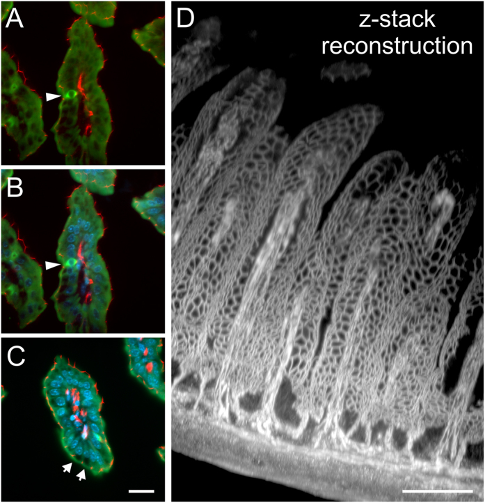
Figure 4. Staining the epithelium.
(A–C) Show cross sections through gut villi stained with the epithelial marker cytokeratin (green) and tight junction associated protein ZO-1 (red, A) in combination with a nuclear DAPI stain (blue; B,C). Cytokeratin is expressed throughout the cytoplasm sparing the nuclei. Some cells show intense cytokeratin stain (arrow heads), whereas goblet cells are devoid of it in the apical half of the cell (arrows in C). The ZO-1 stain allows the localization of the apical junctional complex and thus reveals the epithelial surface organization. Images and reconstruction from a light sheet image stack recorded with a 20xClr Plan-Neofluar 20x/1.0 Corr nd = 1.45 objective, z-steps 2 μm, total z-range 370 μm. Stack subsets displaying an entire villus are shown in Supplementary video 7, and through a goblet cell in Supplementary video 8. (D) Depicts the projection of a partial stack through the entire intestinal wall illustrating the meshwork of the terminal bar complex in the epithelium and capillary endothelium. Due to the high resolution in the z-axis provided by the light sheet microscope the detail of this image can best be appreciated in a fly-around animation shown in Supplementary video 8. Scale bar: 100 μm.

