Figure 2.
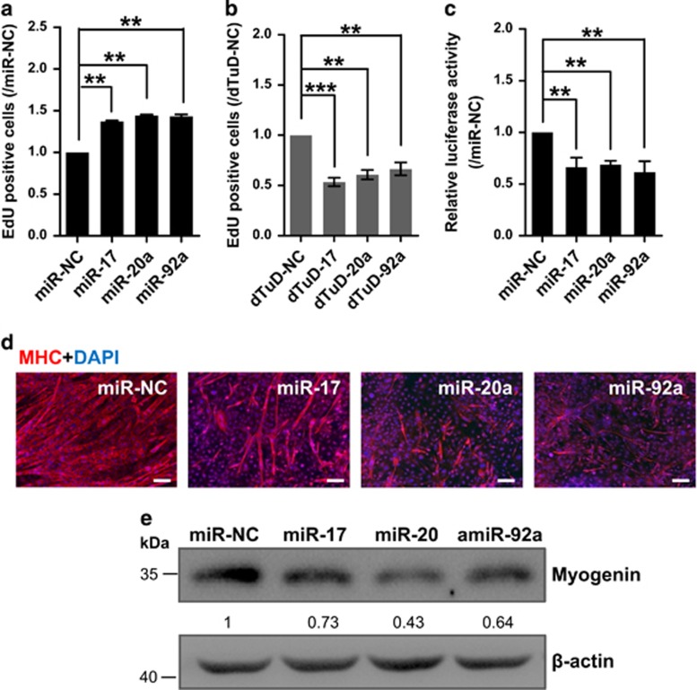
miR-17-92 enhances myoblast proliferation and represses differentiation. (a) Overexpression of miR-17, -20a or -92a enhances the rate of myoblast proliferation. The cell proliferation analysis was performed by EdU incorporation of myoblasts infected with lentiviruses expressing miR-17, -20a or -92a. The cell number of the negative control (miR-NC) was set to 1.0. The error bars depict the means±S.D. of three replicates. **P<0.01. (b) Inhibition of miR-17, -20a or -92a reduces the rate of myoblast proliferation. C2C12 mouse myoblasts were lentivirally infected with the miRNA inhibitors dTuDs targeting miR-17, -20a or -92a (dTuD-17, -20a or -92a) or dTuD-NC. **P<0.01 and ***P<0.001. The rest is as in a. (c) Myogenin promoter luciferase assay demonstrates that miR-17, -20a and -92a reduce the activity of myogenin promoter. C2C12 myoblasts were co-transfected with the pSV40-R.Luc vector, the myogenin promoter luciferase reporter and the vector expressing miR-17, -20a, -92a or NC, and the cells were transferred to DM for 3 days. The luciferase activity of the myoblast transfected with miR-NC was designated as 1.0. The error bars depict the means±S.D. of three replicates. **P<0.01. (d) The miR-17, -20a or -92a mimics repress C2C12 myoblast differentiation. C2C12 myoblasts were transfected with the miRNA mimics as indicated, and the cells were transferred to DM and then stained for MHC (red) and DAPI (blue) at DM4. Scale bar, 100 μm. (e) Western blot shows that the overexpression of miR-17, -20a or -92a reduces myogenin protein expression. C2C12 myoblasts were transfected with miRNA mimics or NC and transferred to DM for 4 days. Beta-actin served as the loading control. The data are presented as the means of the samples from three different cell samples

