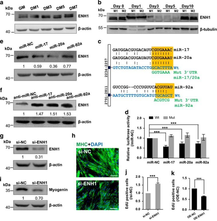
Figure 3.
miR-17, -20a and -92a downregulate ENH1 protein expression by directly targeting its 3′UTR and ENH1 promotes myogenic differentiation but inhibits cell proliferation. (a) ENH1 protein expression during C2C12 myoblast myogenesis. C2C12 mouse myoblasts were cultured in growth medium (GM) and then switched to differentiation medium (DM) for 1–7 days. Beta-actin served as the loading control. (b) ENH1 protein expression in skeletal muscle during regeneration after CTX injury. Beta-tubulin served as the loading control. M1 and M2, mouse 1 and mouse 2. (c) Schematic illustration of the predicted binding sites for miR-17 and -20a (2234-2257) and for miR-92a (2791-2811) in the 3′UTR of ENH1. The wild -type miRNA-binding sites (orange) were modified to complementary sequences (green) to construct the mutated ENH1 3′UTR. (d) ENH1 3′UTR or 3′UTR Mut constructs were co-transfected with the miR-17, -20a or -92a mimics into 293 A cells. The data were normalized to the Renilla luciferase activity. The luciferase activity in cells transfected with miR-NC was set to 1.0. The error bars depict the means±S.D. of three measurements. ***P<0.001. (e) Western blot analysis of ENH1 protein in the C2C12 myoblasts transfected with miR-17, -20a, -92a or NC mimics and (f) with miR-17, -20a, -92a or NC inhibitors. Beta-actin served as the loading control. The data are presented as the means of the samples from three different cell samples. (g) Western blot confirms the efficiency of si-ENH1 on ENH1 protein expression in myoblasts. The rest is as in e. (h and i) si-ENH1 blocks C2C12 myoblast differentiation. C2C12 myoblasts were transfected with si-ENH1 or si-NC and stimulated to differentiate. Immunostaining of MHC (h) and western blot (i) of myogenin showing that si-ENH1 resulted in smaller myotubes and a decrease of myogenin protein at DM4. Scale bar, 200 μm. The rest is as in e. (j and k) ENH1 modulates cell proliferation. C2C12 myoblasts were lentivirally transfected with si-ENH1 or infected with vectors expressing ENH1 and maintained in GM. The EdU incorporation assay was performed for the analysis of cell proliferation. The silencing of ENH1 (j) enhanced EdU incorporation compared with the control (si-NC), whereas the overexpression of ENH1 (k) inhibited cell proliferation. The error bars depict the means±S.D. of three measurements. ***P<0.001

