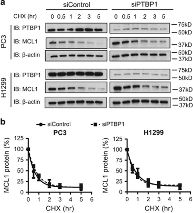
Figure 4.
PTBP1 does not regulate MCL1 protein stability. PC3 and H1299 cells transfected with either siControl or a mixture of two siPTBP1 for 48 h were treated with CHX for time periods as indicated. (a) MCL1 protein levels were assessed by western blotting. (b) Relative MCL1 band intensity in a was quantified and normalized to β-actin. Data were plotted as the percentage of MCL1 protein remaining versus the times of incubation with CHX. The MCL1 protein decay curves were generated using one-phase exponential decay in Graphpad Prism. Each point was presented as mean±S.E.M., n=3

