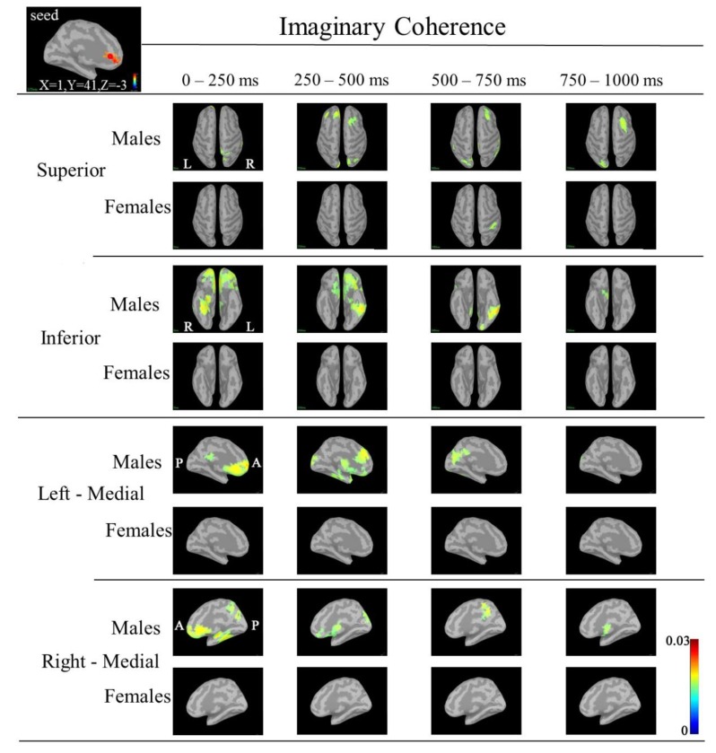
Fig. (7).
IC maps between the seed (left rACC; MNI coordinate: X=1, Y=41, Z=-3) and targets at 30-50 Hz. IC in males was markedly increased in the following targets: bilateral medial prefrontal; right superior parietal, bilateral orbitofrontal, right fusiform regions during 0-250 ms; bilateral frontal, bilateral orbitofrontal (left>right), bilateral occipital, left inferior temporal and left fusiform regions during 250-500 ms; right frontal, left occipital, left cuneus, right isthmus cingulate, left inferior temporal and left fusiform regions during 500-750 ms; and right frontal, right inferior frontal and left occipital regions during 750-1000 ms. IC in females was increased in the supramarginal region during 500-750 ms. (A red circle shows seed point; MNI: Montreal Neurological Institute; IC: imaginary coherence; rACC; rostral anterior cingulate cortex).

