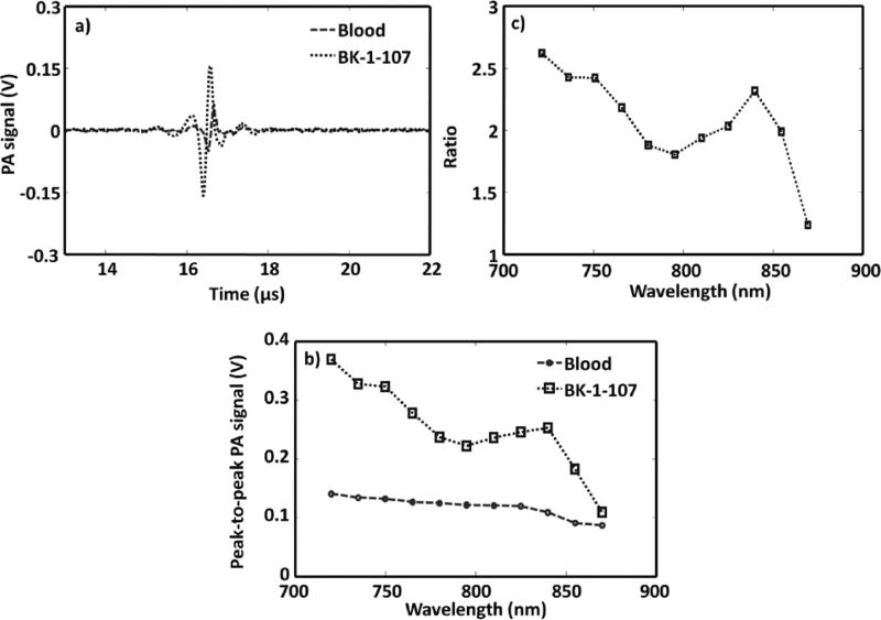
Figure 2.
PA imaging of NIR polymer nanoparticle against blood. a) PA spectroscopy signals generated from a tygon tube (I.D. 250 μm, O.D. 500 μm) filled with NIR-polymer micelle and rat blood. The laser was tuned to 750 nm wavelength. b) PA spectrum of NIR-polymer micelle and rat blood over a 720–870 nm NIR wavelength range. c) Ratio of the peak-to-peak PA signal amplitudes generated from polymeric nanoparticle to those of blood.

