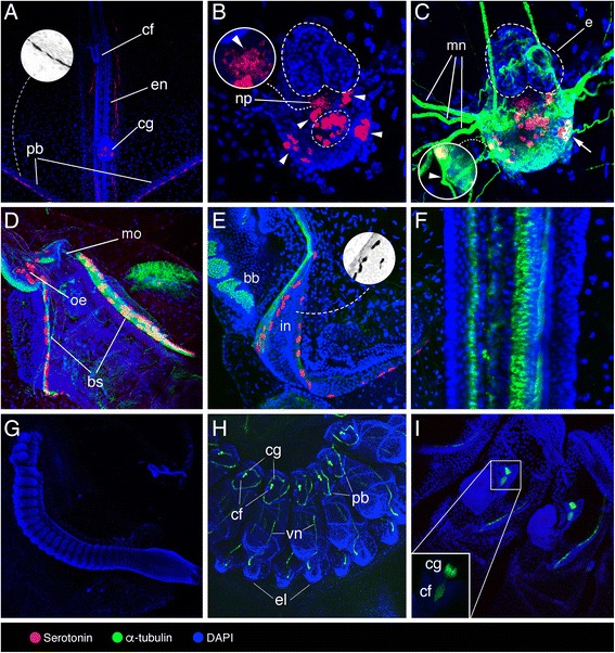
Fig. 2.

Localization of serotonin-like immunoreactivity, acetylated α-tubulin, and DAPI in Thalia democratica. a–f Adult oozooids. g–i Aggregate blastozooids. a General view of the anterior region that contains the ciliated funnel (cf), endostyle (en), cerebral ganglion (cg), and pericoronal bands (pb), with grayscale invert editing to highlight serotonin-like immunoreactive cell shape in the pericoronal bands (inset). b Detail of the cerebral ganglion highlighting peripheral (arrowheads) and central (encircled) serotonin-like immunoreactive cells, and fibres projecting ventrally through the neuropil (arrowhead in the inset). c Detail of the cerebral ganglion highlighting eye (e), neuropil (np) (arrow indicates α-tubulin and serotonin co-labelled neuron), and motor nerves (mn) extending from peripheral serotonergic neurons (arrowhead indicates α-tubulin immunoreactive nerve). d Detail of mouth (mo), oesophagus (oe) and branchial septum (bs). e Magnification of intestine (in) and branchial barrier (bb), with grayscale invert editing to highlight serotonin-like immunoreactive cell shape (inset). f Detail of the endostyle. g General view of early aggregate blastozooids at developmental stage I sensu Brien [39]. h, i Details of aggregate blastozooids at developmental stage II sensu Brien [39] highlighting ciliated funnel (cf), cerebral ganglion (cg), pericoronal bands (pb), visceral nerve (vn), and eleoblast (el)
