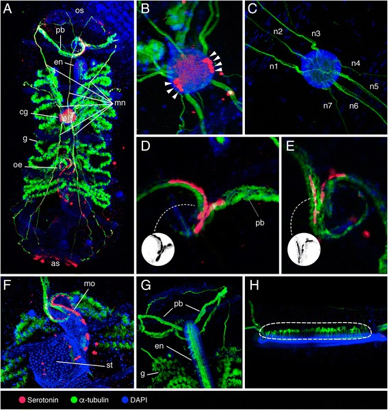
Fig. 4.

Localization of serotonin-like immunoreactivity, acetylated α-tubulin, and DAPI in Doliolina muelleri phorozooid. a Dorsal view of the whole mount phorozooid, highlighting oral siphon (os), pericoronal bands (pb), endostyle (en), motor nerves (mn), cerebral ganglion (cg), gills (g), oesophagus (oe), and atrial siphon (as). b, c Cerebral ganglion with lateral clusters of serotonergic neurons (arrowheads), and motor nerves protruding from it (mn 1–7) at different magnifications. d, e Pericoronal bands (pb), with grayscale invert editing to highlight serotonin-like immunoreactive cell shape (inset). f Initial tract of the digestive system highlighting stomach (st) and serotonergic cells in the mouth (mo). g Anterior part of the specimen highlighting pericoronal bands (pb), endostyle (en), and gills (g). h Lateral view of the endostyle highlighting the long cilia protruding from it (encircled)
