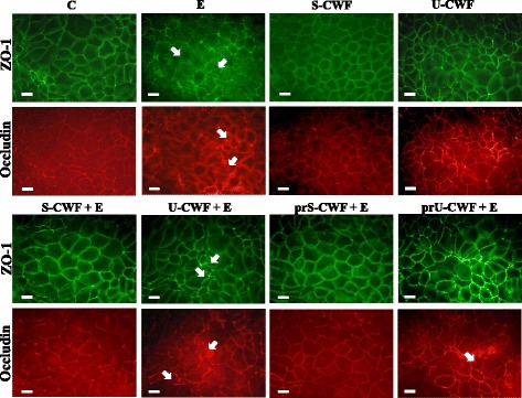
Fig. 1.

S-layer protein-coated or uncoated cell wall fragments differently protect tight junctions (TJs) in F4+ETEC infection. Caco-2/TC7 cells were: untreated (control, C), infected with ETEC (E), treated with S-layer protein-coated cell wall fragments (S-CWF) or uncoated cell wall fragments (U-CWF), co-treated with S-CWF or U-CWF and E (S-CWF + E or U-CWF + E, respectively), pre-treated with S-CWF or U-CWF before F4+ETEC infection (prS-CWF + E or prU-CWF + E, respectively). Cells were labelled with specific primary antibodies for ZO-1 and occludin, followed by FITC- and TRITC- conjugated secondary antibodies for ZO-1 and occludin, respectively. The figure shows the interruption of continuous staining of ZO-1 and occludin, resulting in dissociation of the proteins from membranes in F4+ETEC infected and U-CWF + F4+ETEC treated cells (arrows). Regular localization of TJ proteins is visible in cells pre-treated with S-CWF or co-incubated with S-CWF and F4+ETEC. A delocalization of occludin is present only in few cells pre-treated with U-CWF (arrow). Each figure is representative of three independent immunofluorescence assays (63 × magnification). Bars represent 10 μm
