Figure 2.

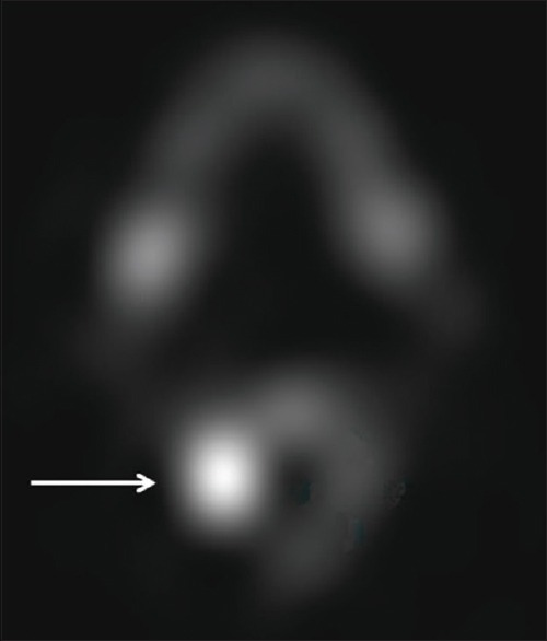
Axial 99mTc-methylene diphosphonate single-photon emission computed tomography image demonstrates focal increased activity (arrow) at the nidus

Axial 99mTc-methylene diphosphonate single-photon emission computed tomography image demonstrates focal increased activity (arrow) at the nidus