Figure 5.

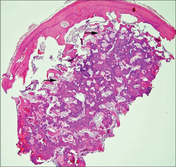
Photomicrograph of the osteoid osteoma, characterized by haphazardly anastomosing bony trabeculae with a sclerotic peripheral rim. The trabeculae are lined by a single layer of osteoblasts (arrows) (H and E)

Photomicrograph of the osteoid osteoma, characterized by haphazardly anastomosing bony trabeculae with a sclerotic peripheral rim. The trabeculae are lined by a single layer of osteoblasts (arrows) (H and E)