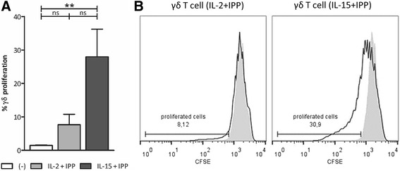
Fig. 1.

Purified γδ T cell stimulation with IL-15 and IPP for 5 days induces their proliferation. a Isolated γδ T cells were stimulated with IL-2+IPP (gray bar) or IL-15+IPP (dark bar) for 5 days. Unstimulated γδ T cells (white bar) were used as negative control. The percentage of proliferated (CFSE-diluted) cells within the viable γδ T cell population was determined by flow cytometry (n = 5) b. Histogram overlays show CFSE dilution and gating of unstimulated γδ T cells (gray-filled area) and γδ T cells exposed to IL-2+IPP or IL-15+IPP (black line) for one representative donor. Friedman test with Dunn’s multiple comparison test. **p < 0.01; ns not significant
