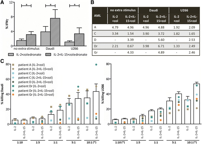
Fig. 6.

Pro-inflammatory and cytolytic γδ T cell capacity after expansion. Percentage IFN-γ-positive γδ T cells after expansion (no extra stimulus) and after an additional 4-h stimulation with Daudi or U266 cells (E:T ratio = 5:1) measured by flow cytometry for a six healthy donors and b five AML patients. c Expanded γδ T cells, with IL2+zoledronate (IL-2+zol) or IL-2+IL-15+zoledronate (IL-2+IL-15+zol), were analyzed by flow cytometry for cytotoxicity against Daudi and U266. Target cell killing was determined by annexin V/PI staining after 4-h incubation at different E:T ratios. Bar graphs (white) represent the mean percentage killing of six healthy donors in three independent experiments. The dots display the proportion of tumor cell killing by γδ T cells of the different patients. Wilcoxon matched-pairs signed rank test. - no value due to low cell number; *p < 0.05
