
Figure 3.
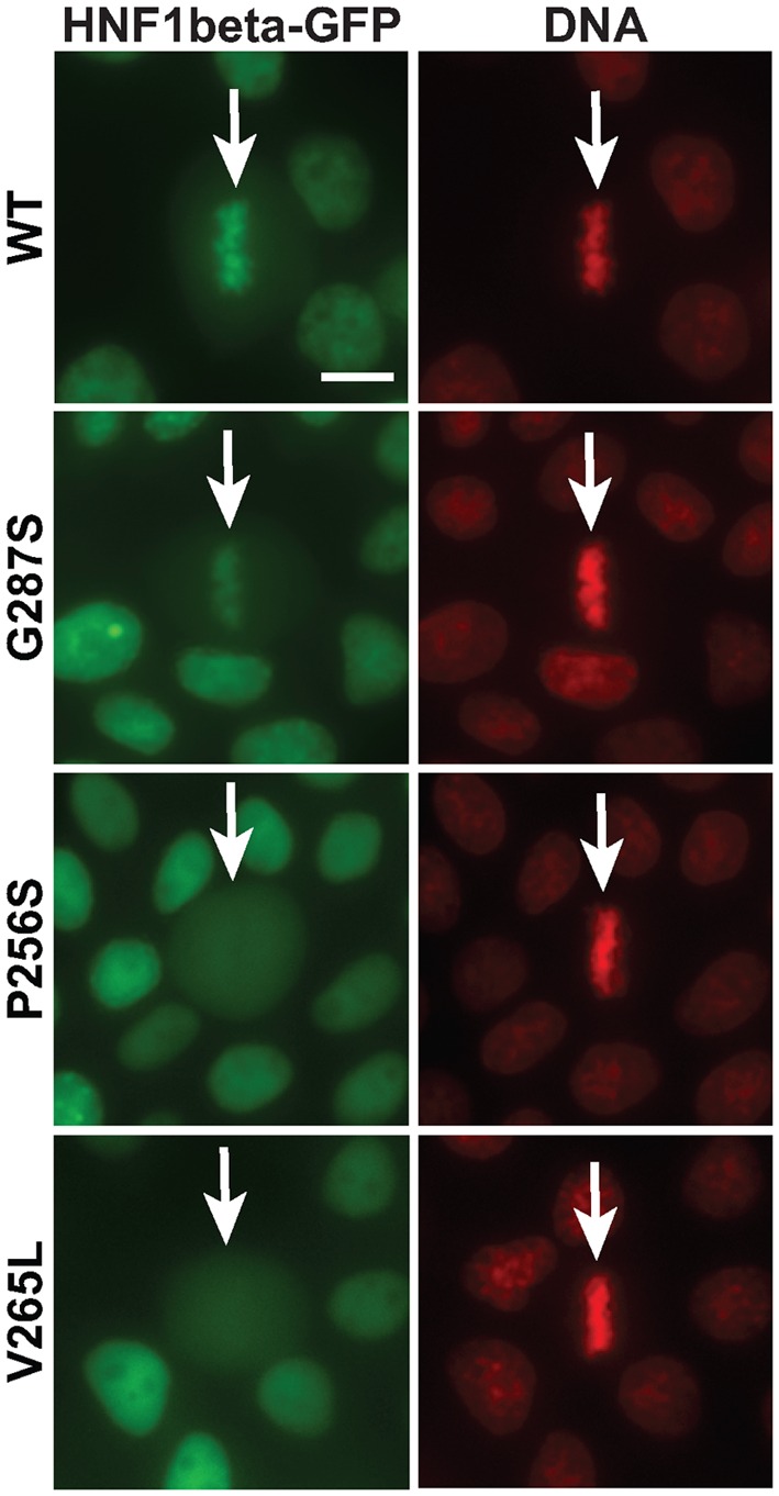
Mitotic chromatin localization defect of MODY mutants. Live imaging of MDCK cells expressing Nter-HNF1β-GFP (WT or MODY mutants) counterstained with Hoechst 33342 (red signal, DNA). White arrows indicate the position of the mitotic cells. Scale bar, 10 μm.