Figure 4.
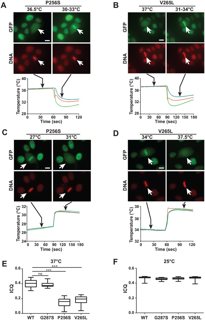
MODY mutations are thermosensitive. (A–D) Effect of abrupt temperature variation on the mitotic chromatin localization of MODY mutations in Nter-HNF1β-GFP fusion protein (green signal) expressed in MDCK cells (red signal, DNA). The temperature in the medium was measured with three independent probes, as shown by the temperature curves. Black arrows indicate the moment in which the picture was taken. P256S and V265L mutants, when grown at restrictive temperature, were dissociated from mitotic chromatin. However, they rapidly re-associated to mitotic chromatin upon sudden temperature drop (panels A and B). Conversely, they rapidly dissociated from mitotic chromatin when temperature was rapidly raised (panels C and D). White arrows indicate the position of the mitotic cells. Scale bars, 10 μm. (E and F) Quantification of the ‘Intensity Correlation Quotient’ (ICQ) between Hoechst and GFP signals at 37°C (panel E) or at room temperature (panel F), in mitotic MDCK cells stably expressing Nter-HNF1β-GFP WT or mutants. WT 37°C, n = 34; G287S 37°C, n = 32; P256S 37°C, n = 32; V265L 37°C, n = 35; WT 25°C, n = 30; G287S 25°C, n = 34; P256S 25°C, n = 26; V265L 25°C, n = 31. Statistical significance was determined by Kruskal–Wallis test followed by Dunn's multiple comparisons test. ***P < 0.0001.

