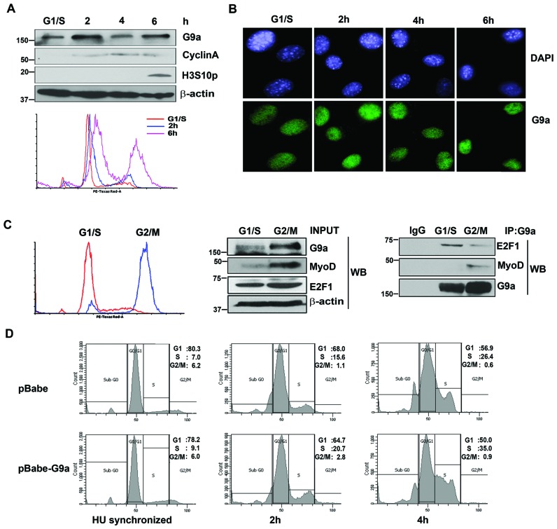
Figure 6.
G9a associates with E2F1 and MyoD in distinct phases of the cell cycle. (A) C2C12 cells were synchronized at G1/S boundary using HU. Transition into the S phase (2 and 4 hr culture in growth media after HU removal) and mitosis (6 hr culture in growth media after HU removal) was checked using CyclinA, as a marker of the S phase, and Histone H3 serine 10 phosphorylation (H3S10p) as a mitotic marker. G9a expression was checked using western blot. β-actin was used as an internal control. Synchronization of cells with HU was also confirmed by flow cytometry using PI staining (lower panel). (B) HU synchronized C2C12 cells were released and fixed 2, 4 and 6 hr after HU removal and immunostained with anti-G9a antibody (green). Nuclei were counterstained with DAPI (blue). (C) C2C12 cells were synchronized at G1/S and G2/M boundary using HU and nocodozole respectively. Synchronization was examined by flow cytometry (left panel). The expression of G9a, MyoD and E2F1 in lysates is shown (middle panel). Endogenous G9a was immunoprecipitated from synchronized cells and tested for association with E2F1 and MyoD respectively (right panel). The interaction of G9a with E2F1 is higher at G1/S phase, and with MyoD at the G2/M phase. (D) Flow cytometric analysis of pBabe and pBabe-G9a cells synchronized at G1/S boundary using HU and released into growth media for 2hr and 4hr. The results are representative of at least two independent experiments.

