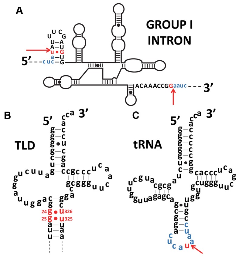
Figure 6.

Similarity of the positions of G:U mismatches in the tRNA intron and in tmRNA. (A) Secondary structure of the Azoarcus group I intron. Exon sequences are in lower case and blue, while introns are in upper case letters, with red arrows indicating the splice boundaries. The conserved G-U mismatch necessary for self-splicing and the guanine partner are red. (B) Secondary structure of the Escherichia coli tRNA-like domain (TLD) of tmRNA. The conserved G-U mismatches in the TLD are red. A similarity in position between the G-U mismatch of the tRNA intron and the TLD is noticeable. (C) Secondary structure of a tRNAIle (CAU) from Azoarcus. The red arrow indicates the insertion site for the introns shown in (C). The exon sequence common to figures (A) and (C) is blue. Mismatches are signaled by dots.
