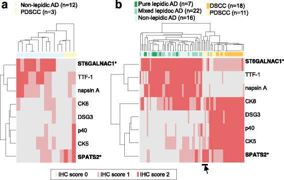
Fig. 4.

The results of IHC with the novel and known markers. (a) The presence of the known markers and the candidate markers was examined by IHC of carcinoma tissues of non-lepidic AD and PDSCC obtained from the same patients evaluated in the CAGE analysis. The staining patterns are scored (IHC score 0, 1, and 2) as described in the METHODS section, and the scores are visualized as heatmaps, where the tissues and markers are clustered based on the IHC scores. (b) Equivalent heatmaps based on the results of an independent group of patients, consisting of pure lepidic AD and mixed lepidic AD, DSCC, as well as non-lepidic AD and PDSCC
