
Figure 4. PKC is an upstream mediator of STAT3α and GSK3β phosphorylation induced by TPA.
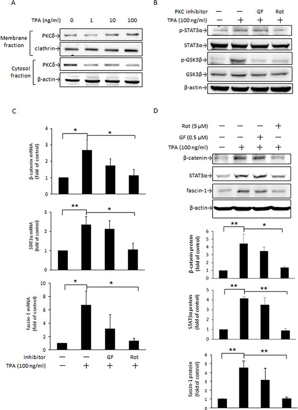
MCF-7 cells were treated with various concentrations of TPA for 30 min and PKCδ in the plasma membrane and cytosolic fractions were determined (A). Cells were pretreated with or without 0.5 μM nonselective PKC inhibitor GF109203X (GF) or 5 μM PKCδ-specific inhibitor rottlerin (Rot) for 1 h followed by incubation with 100 ng/ml of TPA for another 30 min. STAT3α and GSK3β phosphorylation were measured (B). β-Catenin, STAT3α, and fascin-1 mRNA (C) and protein (D) levels were determined after 18 h and 24 h with TPA treatment, respectively. One representative experiment out of three independent experiments is shown. Mean ± SD, n = 3. *p < 0.05 and **p < 0.01.