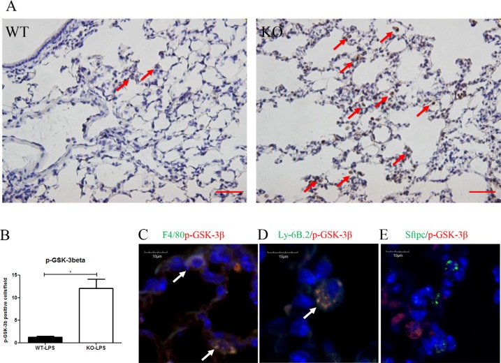
Figure 6.
(A, B) Immunostaining for p-GSK-3β showed the number of p-GSK-3β+ cells (brown) were significantly higher at Day 2 after LPS treatment in Xb130 KO mice compared to WT mice. Scale Bars = 50 μm. p-GSK-3β was mainly stained in neutrophils and macrophages (C, D) but not in alveolar type II cells (E). *p < 0.05, WT = Wild type, KO = Xb130 knockout.

