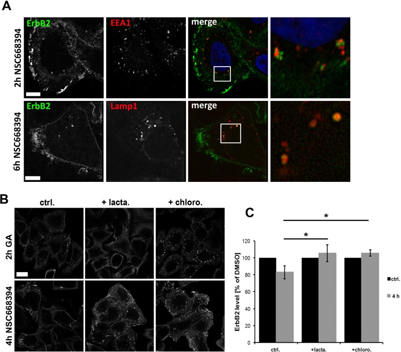
Figure 6. Analysis of intracellular sorting of ErbB2 by confocal microscopy.
(A) ErbB2 localization with EEA1 and Lamp1 after ERM inhibition. Cells were treated for 2 h or 6 h with NSC668394 and subsequently fixed and stained for ErbB2 and EEA1 or Lamp1. ERM inhibition leads to ErbB2 internalization and association to the endosomal marker EEA1 at early time points and association Lamp1-possitive vesicles at later time points. Scale bars: 10 μm. (B) Cells were pretreated for 2 h with 10 μM of lactacystin or chloroquine, followed by a 2 h treatment with 3 μM geldanamycin (GA) or 4 h with 30 μM of NSC668394. Afterwards, cells were fixed and stained for ErbB2. Lactacystin inhibits the uptake of ErbB2 triggered by GA but not by NSC668394. Chloroquine pretreatment leads in GA and NSC668394 treated cells to an intracellular accumulation of ErbB2 to vesicular structures. Scale bar: 10 μm. (C) Quantification of ErbB2 protein levels after lactacystin and chloroquine treatment. Cells were treated as described in (B), lysed and analyzed by SDS-PAGE. Treatment of cells with lactacystin or chloroquine inhibits degradation of internalized ErbB upon ERM inhibition by NSC668394. Data is shown as mean +/− SEM (*P < 0.05).

