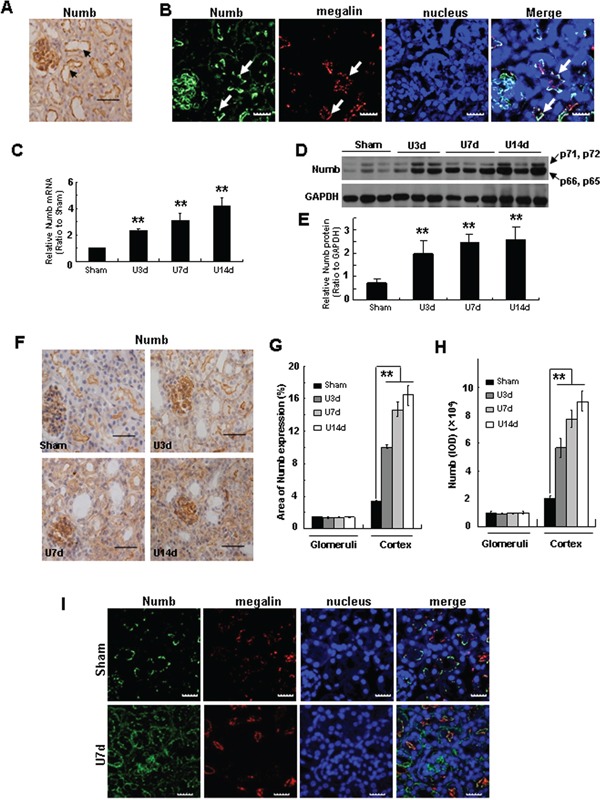
Figure 1. Numb expression is induced in TECs after obstructive injury.

A. Immunohistochemistry staining by using anti-Numb antibody shows the abundance and distribution of Numb protein in the kidney of C57BL/6J mice at the age of 8-12 weeks (see arrows). Bar=50μm. B. Immunofluorescence staining shows the co-localization of Numb (green) and megalin (red) in proximal tubules (see arrows). Nuclei were stained with DAPI (blue). Images were taken by confocal microscopy. Bar=20μm. C. Real time-PCR shows the level of Numb mRNA in injured kidney was increased in a time-dependent manner after UUO. C57BL/6J mice were subjected to UUO, and kidney tissues were collected at different time points after surgery as indicated. Relative Numb mRNA levels were expressed as fold induction over sham controls after normalization with GAPDH. D. Western blot analysis shows the induction of Numb protein in fibrotic kidney induced by UUO. E. Graphic representation of relative protein level of Numb normalized to GADPH. F. Immunohistochemistry staining shows the expression and distribution of Numb in the kidney at day 3, 7 and 14 after UUO. Bar=50μm. Quantification of the percentage of the Numb-positive area G. and the IOD of Numb H. in kidney sections. I. Immunofluorescence staining shows the co-staining of Numb (green) with megalin (red) in UUO. Bar=20μm. Data are expressed as mean±SD, n=6. **p<0.01 versus sham.
