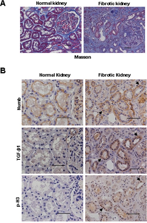
Figure 8. Increased expression of Numb in human fibrotic kidney.

A. Representative micrographs show the collagen deposition detected by Masson trichrome staining in human normal and fibrotic kidneys. Bar=50μm. B. Representative images of the immunohistochemical staining of Numb, TGF-β1 and p-H3 in sequential sections of renal biopsies from patients with renal tubulointerstitial fibrosis. Normal renal tissues adjacent to tumor (renal cell carcinoma) were used as normal control. Arrows indicate the co-localization of Numb, TGF-β1 and p-H3 in the injured tubules. Bar=50μm.
