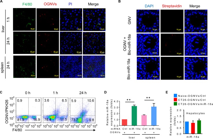
Figure 1. OGNV-mediated delivery of miRNA is taken up by mouse Kupffer cells in vivo.
(A) PKH26-labeled (red) OGNVs located in liver Kupffer cells (F4/80+, green), not in spleen macrophages (F4/80+, green) from BALB/c mice are visualized with confocal microscopy, assessed 1 h and 24 h after intravenous injection. (B) Analysis of Alexa Fluor fluorescent streptavidin conjugates with confocal microscope, assessed 24 h after intravenous injection of OGNVs alone, OGNVs with biotin-conjugated miR-18a (bio-miR-18a), or bio-miR-18a alone. (C) Frequency of F4/80+ cells and PKH26-labled OGNVs in the liver from BALB/c mice assessed using flow cytometry. Numbers in quadrants indicate percent cells in each. (D) Quantification of miR-18a level in leukocytes from BALB/c mouse liver and spleen assessed 24 h after intravenous injection of OGNVs with miR-18a by quantitative real-time PCR (qPCR). *P < 0.05 and **P < 0.01 (two-tailed t-test). Data are representative of three independent experiments (error bars, S.E.M.). (E) Expression of miR-18a in hepatocytes from naive BALB/c mice, CT26 liver metastasis mice with OGNVs/Ctrl or OGNVs/miR-18a treatment assessed by quantitative real-time PCR (qPCR).

