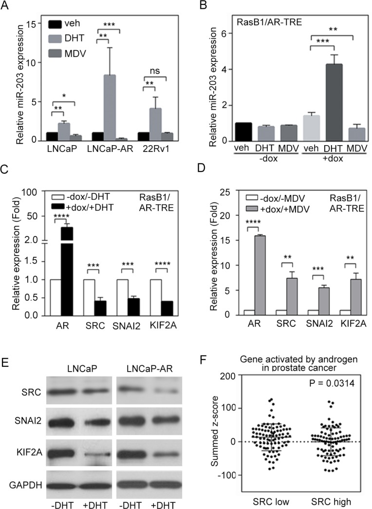
Figure 3. Androgen receptor (AR) signaling inversely regulates SRC and miR-203 levels.
(A) Relative miR-203 expression in LNCaP, LNCaP-AR, and 22Rv1 cells in the presence of vehicle, dihydrotestosterone (DHT), or MDV3100. The data are presented as the mean ± SEM, n = 3. *p < 0.05, **p < 0.01, ***p < 0.001. (B) Relative miR-203 expression in AR-TRE-transfected RasB1 cells with or without doxycycline (dox) induction in the presence of vehicle, DHT, or MDV3100. The data are presented as the mean ± SEM, n = 3. **p < 0.01, ***p < 0.001. (C and D) Relative AR, SRC, SNAI2, and KIF2A mRNA expression in AR-TRE-transfected RasB1 cells with or without doxycycline induction in the presence or absence of DHT (C) or MDV3100 (D). The data are presented as the mean ± SEM, n = 3. **p < 0.01, ***p < 0.001, ****p < 0.0001. (E) Relative SRC, SNAI2, and KIF2A protein expression in LNCaP and LNCaP-AR cells in the presence or absence of DHT. (F) Mean summed z-scores for AR signaling-responsive gene signatures in the Taylor PCa dataset (n = 111), showing that patients with high SRC expression had lower expression of genes that are activated by AR signaling. Statistical significance was determined by Student's t-test.

