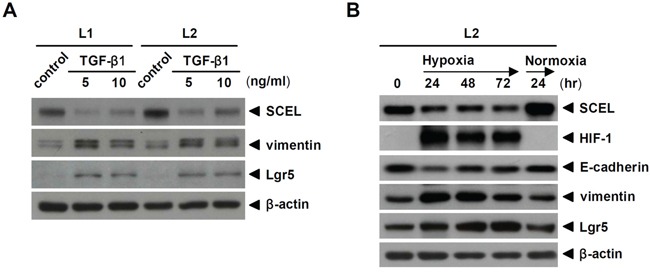
Figure 5. TGF-β1 and hypoxia inhibit SCEL expression.

A. L1 and L2 were treated with 5 and 10 ng/mL TGF-β1 individually for 24hr. The expression of SCEL, vimentin, and Lgr5 was determined using western blot. B. L2 was cultured in hypoxic condition for 3 days and then restored to normoxic condition for 1 day. The expression of SCEL, HIF-1, E-cadherin, vimentin, and Lgr5 was determined using western blot. β-actin served as loading control.
