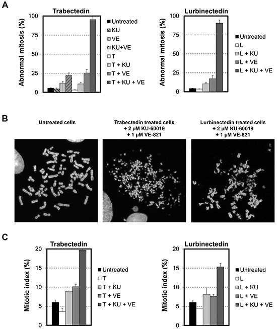
Figure 7. Influence of the combination of checkpoint abrogators on DSBs repair.

A. HeLa cells were exposed to 1 nM trabectedin (left panel, T) or lurbinectedin (right panel, L) for 1 hour in the absence (white columns) or presence of 2 μM KU-60019 (+ KU, light grey columns), 1 μM VE-821 (+ VE, medium grey columns) or a combination of 2 μM KU-600019 and 1 μM VE-821 (+ KU + VE, dark grey columns). This was followed by 24 hours post-incubation in the absence (white columns) or presence of 2 μM KU-60019 (+ KU, light grey columns), 1 μM VE-821 (+ VE, medium grey columns) or a combination of 2 μM KU-600019 and 1 μM VE-821 (KU + VE, dark grey columns). Cells were then processed for karyotype analysis. Untreated cells were used as a negative control (black columns). The left panel shows the influence on HeLa cells of 2 μM KU-60019 (KU, light grey dashed column), 1 μM VE-821 (VE, medium grey dashed column) or a combination of 2 μM KU-600019 and 1 μM VE-821 (KU + VE, dark grey dashed column) when they were given in the absence of ETs. Data are represented as mean +/− SD. B. Typical metaphase in untreated HeLa cells and cells treated for 1 hour with 1 nM of either trabectedin or lurbinectedin combined with a combination of 2 μM KU-600019 and 1 μM VE-821 and post-incubated for 24 hours in the presence of a combination of 2 μM KU-600019 and 1 μM VE-821. C. The mitotic index was determined on the microscopy slides used for karyotype analysis. Data are expressed as mean +/− SD.
