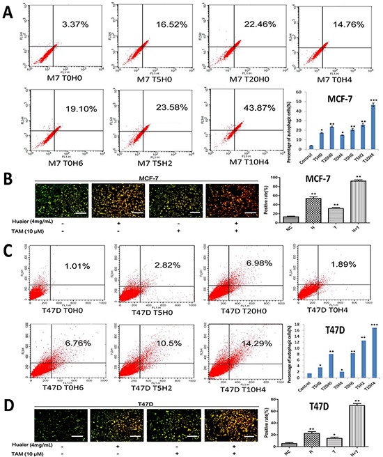
Figure 3. Huaier extract synergizes with tamoxifen to induce autophagy in ER-positive breast cancer cells.

MCF-7 A. and T47D C. cells were labeled with Acridine orange (AO) after the indicated treatments and quantified using flow cytometry. FL1-H indicates green color intensity (cytoplasm and nucleus), whereas FL3-H shows red color intensity (AVO). Cells in up quadrants were considered AVO-positive. Treated MCF-7 B. and T47D D. cells were stained with AO and examined under a fluorescence microscope. Bars, 50 μm. All of the experiments were performed in triplicate and data are presented as the mean ± SD of three separate experiments (*p < 0.05; **p < 0.01; ***p < 0.001 vs. the control group).
