
Figure 6. The effect of miR-124-1 on tumor formation in a nude mice xenograft model.
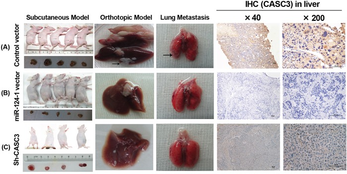
Nude mice were injected subcutaneously in opposite flanks with 5×10⁶ control lentiviral vector-infected cells and miR-124-1 vector-infected cells. After 2 weeks, the mice were sacrificed when the tumors reached 1.0 cm in diameter and the subcutaneous tumors were cut into 1.0 mm3 sections, which were then inserted into the livers of another 10 nude mice. The mice were followed for 30 days and then killed by cervical dislocation. Livers and lungs were resected and imaged with a high-definition digital camera. Each group was composed of 5 mice. The weight of the tumors in the two groups was compared using the Student's t-test. A. control lentiviral vector groups; B. miR-124-1 vector groups; C. Sh-CASC3 groups.