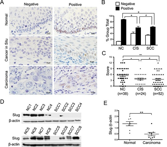
Figure 1. Expression of slug in normal cervix samples and various cervical lesions.

(A) Immunohistochemical (IHC) detection of Slug in normal cervix, cancer in situ and carcinoma samples; original magnification, 1000×. (B) Slug staining is classified into 2 categories (negative and positive), and the bar chart shows the percentage of each group (38 normal cervix specimens, 24 carcinoma in situ specimens, and 52 invasion carcinoma tissue specimens). (C) The scatter plot shows the immunoreactivity scores (IHC) obtained for the staining of Slug in normal cervix, cervical cancer in situ and invasive cervical cancer samples (points represent the IHC score per specimen, and one-way ANOVA was performed). (D) The expression of Slug in normal cervix (NC) and squamous cervical carcinoma (SCC) samples was detected using western blotting. (E) The relative expression of Slug in each normal cervix tissue (n = 8) and squamous cervical carcinoma tissue sample (n = 8) is shown. The data shown are the ratios of the Slug/β-actin of each specimen and the means ± standard error of the NC and SCC groups (triangles represent relative Slug expression). Values are shown as the mean ± SD, *p < 0.05, **p < 0.01.
