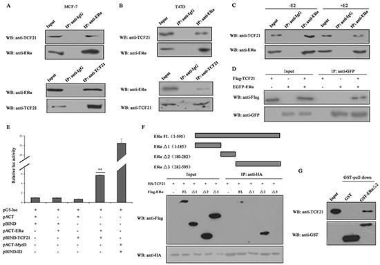
Figure 1. Interaction between TCF21 and ERα.

A–B. MCF-7 and T47D cells were subjected to IP with anti-ERα antibody followed by Western blot with anti-TCF21 and anti-ERα antibodies or vice versa. IP carried out with anti-IgG antibody was used as control. C. MCF-7 cells treated with or without E2 were subjected to IP using anti-ERα antibody followed by Western blot with anti-TCF21 antibody. IP carried out with anti-IgG antibody was used as control. D. HEK 293T cells transfected with Flag-TCF21 only, EGFP-ERα only, or with Flag-TCF21 plus EGFP-ERα were subjected to IP using anti-GFP antibody followed by Western blot with anti-Flag antibody. E. Interaction between TCF21 and ERα as demonstrated by mammalian two hybrid system. TCF21 and ERα were expressed from pBIND-TCF21 and pACT-ERα, respectively, whereas the empty vectors pACT and pBIND were used as controls, as indicated by the pG5-luc reporter in MCF-7 cells. Cells transfected with pBIND-ID and pACT-MyoD were used as positive control. Luciferase activity was measured 36 h after transfection. The luc activity level of cells transfected with pG5-luc, pACT and pBIND was set to 1. Data are the means ± S.Ds of three experiments. ‘**’ indicates significantly different from cells transfected with pACT and pBIND at the P <0.01 level. F. HEK 293T cells were transfected with HA-TCF21 and Flag-tagged full-length ERα (FL), ERα Δ1, ERα Δ2, or ERα Δ3. The cells were subjected to IP using anti-IgG or anti-HA antibody followed by Western blot with anti-Flag antibody. G. Interaction between TCF21 and ERα Δ2 in vitro. Purified His-TCF21 was incubated with immobilized GST- ERα Δ2 or GST alone. The bound proteins were subjected to Western blot assay. All experiments were repeated at least three times. Data are the mean ± SDs of three independent experiments.
