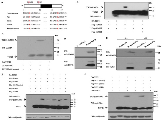
Figure 3. Sumoylation of TCF21.

A. Schematic representation of the primary structures of TCF21 and the predicted sumoylation sites along with the structures of TCF21 in different species. B. HEK 293T cells transfected with HA-TCF21 and Flag-SUMO1, 2 or 3 were subjected to Western blot with anti-HA antibody. C. HEK 293T cells co-transfected with GFP-SUMO1 or GFP-SUMO1 mutant and HA-TCF21 were subjected to Western blot with anti-HA antibody. D. HEK 293T cells were subjected to IP using anti-TCF21 antibody or anti-IgG antibody followed by Western blot using anti-SUMO1 or anti-TCF21 antibody. E. MCF-7 cells treated with or without E2 were subjected to IP with anti-TCF21 antibody followed by Western blot with anti-SUMO1 antibody. F. HEK 293T cells were transfected with different combinations of constructs as indicated, followed by Western blot with anti-HA antibody. No NEM was added in the cell extracts. G. HEK 293T cells were transfected with Flag-tagged wild-type or mutant TCF21(K24R, K65R or 2KR) and GFP-SUMO1, and individual cell extracts were subjected to Western blot with anti-Flag antibody. All experiments were repeated at least three times.
