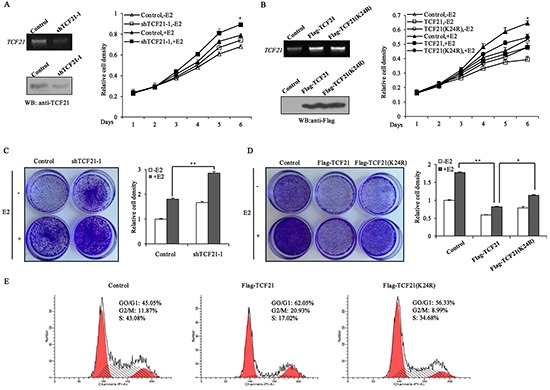
Figure 6. Sumoylation of TCF21 inhibits the growth of breast cancer cell lines.

A. MCF-7 cells were stably transfected with control vector, or shTCF21-1. MTT assay was conducted either with or without pre-treatment of the cells with 10 nM E2 for the indicated times. The mRNA and protein levels of TCF21 were examined by PCR and Western blot assay. B. MCF-7 cells were stably transfected with control vector, Flag-TCF21or Flag-TCF21(K24R). MTT assay was conducted either with or without pre-treatment of the cells with 10 nM E2 for the indicated times. The mRNA and protein levels of TCF21 were examined by PCR and Western blot assay. C. MCF-7 cells stably transfected with control vector, or shTCF21-1 were stained with crystal violet after 8 days of growth (left panel), and the corresponding quantitative analyses are shown on the right panel. For comparison, the number of control cells was set to 1. ‘**’ indicates significantly different from cells transfected with shTCF21-1 at the P<0.01 level. D. MCF-7 cells stably transfected with control vector, Flag-TCF21or Flag-TCF21(K24R) were stained with crystal violet after 8 days of growth (left panel), and the corresponding quantitative analyses are shown on the right panel. For comparison, the number of control cells was set to 1. ‘**’ indicates significantly different from cells transfected with Flag-TCF21 at the P<0.01 level. ‘*’ indicates significantly different from cells transfected with Flag-TCF21 (K24R) at the P<0.05 level. E. MCF-7 cells were transfected with control vector, or with Flag-tagged wild-type or mutant TCF21(K24R). The cell-cycle distribution of MCF-7 cells was examined by flow cytometry analysis after 16 h of growth in presence of 10 nM E2. All experiments were repeated at least three times. Data are the means ± SDs of three independent experiments.
