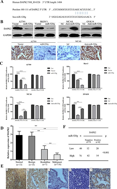
Figure 5. miR-520g directly targets DAPK2 in EOC cells and miR-520g expression was inversely associated with DAPK2 in EOC tissues.

(A) TargetScan predictions showed that miR-520g directly binds DAPK2. (B) Western blotting and tumor xenograft IHC staining showed that miR-520g inhibited DAPK2 post-transcriptionally. (C) Luciferase-dual reporter activity assay showed that miR-520g repressed DAPK2 expression in wild type but not mutant EOC cells. Luciferase-dual reporter vector (Luc) was the control (*p < 0.05, **p < 0.001, ***p < 0.0001). (D) Relative DAPK2 mRNA expression in normal (n = 13), benign (n = 15), borderline (n = 7) and EOC (n = 116) tissues (**p < 0.001). (E) DAPK2 IHC staining in normal, benign, borderline and EOC tissues: E1, strong DAPK2 staining in normal tissue; E2, moderate intensity staining in benign tissue; E3, weak intensity staining in borderline tissue; E4, negative staining in EOC tissue with high miR-520g expression; E5, strong staining in EOC tissue with low miR-520g expression (magnification × 200). (F) miR-520g was negatively correlated with DAPK2 expression in EOC tissues (**p < 0.001).
