
Figure 1. Expression of MIA and STOX2 in human OSCC cases.
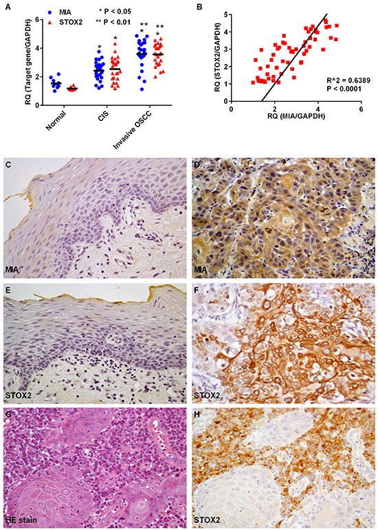
A. Gene expression analysis using cDNA obtained by laser capture microdissection (LCM) in normal epithelium of the oral cavity and oral squamus cell carcinoma (OSCC). Levels of MIA and STOX2 expression in carcinoma in situ (CIS) (P < 0.05) and invasive OSCC (P < 0.01) cases were high compared with that in the normal oral mucosa. Further, MIA and STOX2 expression in patients with invasive OSCCs were higher than in patients with CIS (P < 0.05). B. Overexpression of STOX2 was significantly associated with upregulation of MIA in OSCCs (P < 0.0001). C–H. Immunostaining of MIA and STOX2 in human OSCC cases. Weak and/or no expression of MIA (C) and STOX2 (E) were found in tumor free oral mucosa. Expression of MIA (D) and STOX2 (F) were observed in OSCCs. (G) Stromal plasma cells surrounding the OSCC cell nest. (H) Immunostaining of STOX2 was found in stromal plasma cells. Original magnification was 400×. HE; hematoxylin and eosin