
Figure 3. Expression of the novel HLA-G regulating miR-548q and miR-628-5p in vitro and in vivo.
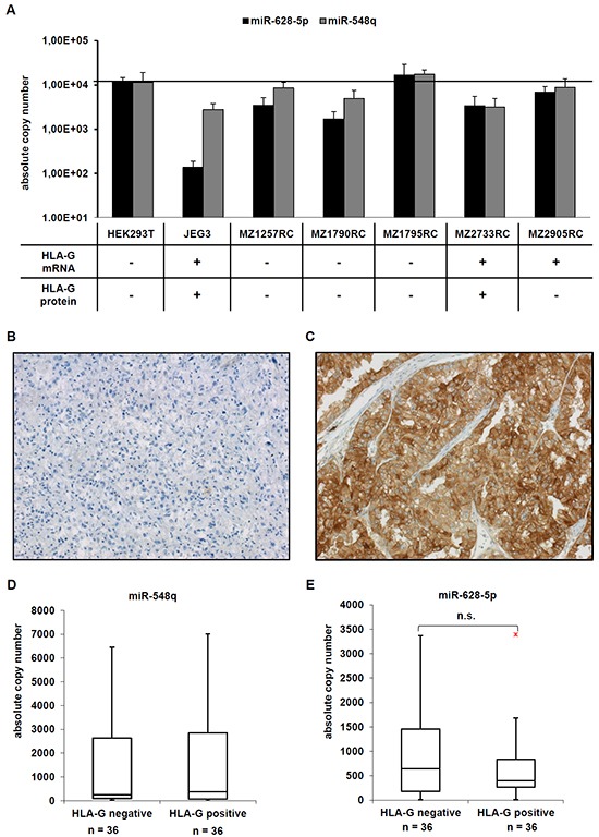
A. A set of five RCC cell lines, the HEK293T cell line and as HLA-G-positive control the choriocarcinoma cell line JEG-3 were analyzed for the expression of HLA-G, miR-548q and miR-628-5p as described in Material and Methods. The HLA-G expression of the cell lines analysed has already been reported in [8] and summarized in the table below the bar diagram. B, C. Two representative immunohistochemical stainings of a HLA-G negative (B) and a HLA-G positive (C) RCC lesion from a tissue microarray consisting of > 450 RCC lesions are shown. D, E. The expression of miR-548q (D) and miR-628-5p (E) was determined in 36 HLA-G negative and 36 HLA-G positive RCC lesions by qPCR and results are visualized in Box-Whisker-Plots. The red dot in Figure 3E is an outlayer.