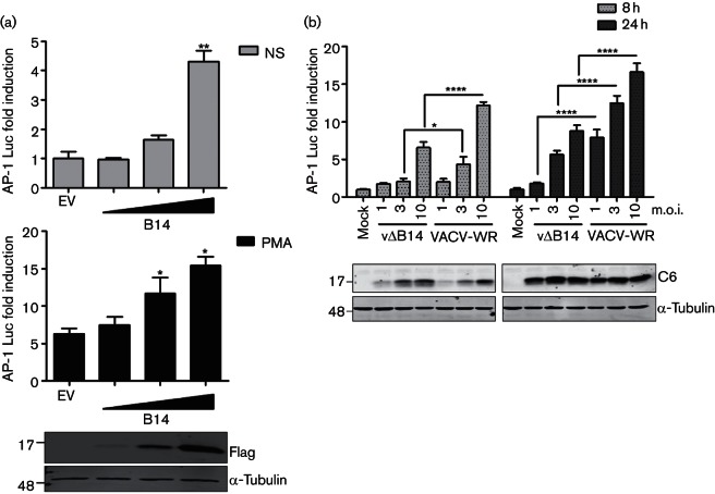
Fig. 2.
B14 stimulates AP-1 in a dose-dependent manner following transfection and infection. (a) HeLa cells were co-transfected in triplicate with an AP-1 luciferase reporter, a plasmid expressing renilla luciferase and increasing amounts of the B14 vector or the empty vector (EV). Twenty-four hours later, the cells were stimulated for 24 h with PMA (bottom graph) or left non-stimulated (NS) (top graph). (b) Reporter gene assay in HeLa cells mock-infected or infected for 8 or 24 h with VACV-WR (wild-type) or B14 deletion (vΔB14) viruses, using the indicated multiplicity of infection (m.o.i.). The luminescence of each sample was measured and normalized to that of EV non-stimulated(NS) (a) or mock-infected cells (b) to give the fold induction. Data are shown as the mean±sd and are representative of three experiments. Statistical analysis was by unpaired Student’s t-test (*P<0.05, **P<0.01, ****P<0.0001) in comparison to EV control, either non-stimulated or PMA-stimulated (a) or between vΔB14 and VACV-WR (b). The panels below each graph show protein expression controls by immunoblotting; molecular masses (in kDa) are indicated on the left.

