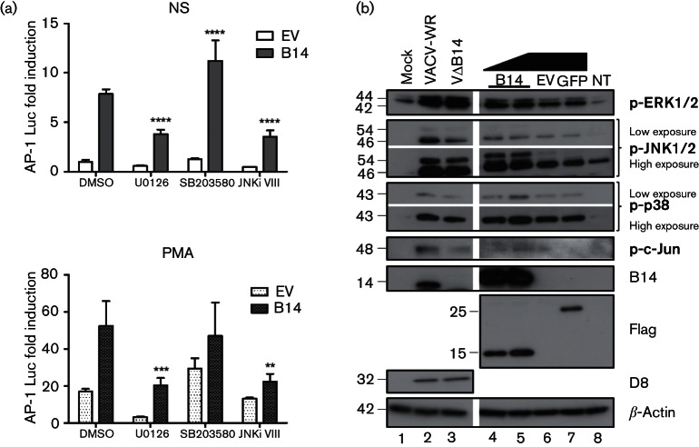
Fig. 3.
Contribution of B14 to MAPK activation (a) HeLa cells were co-transfected in triplicate with an AP-1 luciferase reporter, a renilla luciferase reporter and B14 vectors. After 24 h, cells were treated with U0126 (MEK/ERK inhibitor), SB203580 (p38 MAPK inhibitor), JNK inhibitor VIII (JNK1/2 inhibitor) or DMSO, and stimulated for 24 h with PMA (10 ng ml−1) or left non-stimulated (NS). The luminescence of each sample was measured and normalized to that of the non-stimulated control. Data are shown as the mean±sd and are representative of three experiments. Statistical analysis was by Student’s t-test (**P<0.01, ***P<0.001, ****P<0.0001). (b) HeLa cells were mock-infected or infected with VACV-WR or vΔB14 (lanes 1, 2 and 3, respectively) for 12 h (5 p.f.u. per cell). In parallel, cells were transfected with the B14 (1 or 2 µg), GFP or empty (EV) vectors or left non-transfected (NT) for 24 h (lanes 4, 5, 7, 6 and 8, respectively). Cells were harvested and lysates were subjected to immunoblotting for the proteins shown; molecular masses (in kDa) are indicated on the left.

