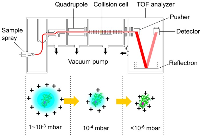
Figure 2.
Electrospray ionization mass spectrometry (ESI-MS). A diagram of the mass spectrometer and a schematic of the electrospray-ionization process are shown. A protein–ligand complex is ionized by electrospray ionization followed by injection into a spectrometer. Under proper vacuum conditions, the solvent molecules gradually dissociate from the complex without the disruption of non-covalent interactions when the protein molecule passes through the low-vacuum chambers. Finally, the mass of the complex is measured under high vacuum by a time-of-flight (TOF) mass spectrometer (modified from [29]; https://www.jstage.jst.go.jp/article/biophys/55/5/55_270/_article/-char/ja/).

