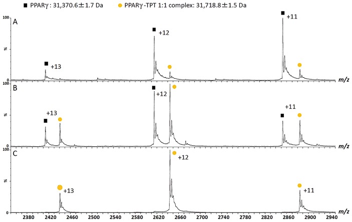
Figure 3.
Mass spectra of the PPARγ–ligand binding domain complex with triphenyltin (TPT) under non-denaturing conditions. The PPARγ–ligand binding domain forms a complex with TPT in a 1:1 molar ratio (A–C). The mass patterns after the addition of aliquots of formic acid (A, 3%; B, 1%; C, 0%) to the complex indicate that the dissociation of the interaction is caused by the unfolding of the PPARγ–ligand binding domain (refer to [7]).

