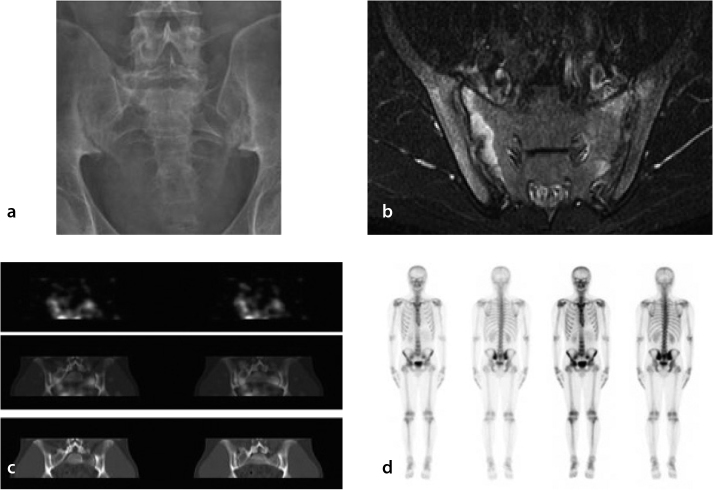
Figure 1.
a–d. Composite images from a patient with AS for comparing various imaging modalities. The subunits are plain X-ray in panel (a), MRI in panel (b), glucosamine scan in panel (c), and bone scan in panel (d). On plain X-ray (panel A), there is sclerosis in both SI joints with erosive changes present. The joint spaces did not appear to be widened. Radiographically, these findings were consistent with Grade 2 sacroiliitis. On the axial short-TI inversion recovery (T1 STIR) MRI image (panel B), there is edema on both sides of the SI joints (right greater than left), which is associated with enhancement suggestive of active inflammatory change. The SI joints were slightly widened with subtle erosive changes bilaterally. There was also minor thickening and enhancement of the synovium. On the 99mTc-glucosamine scans (panel C), diffuse increased tracer uptake was noted in the vicinity of each SI joint. On the early-phase images of the nuclear bone scan there was hyperemia in the SI regions (panel D). On the planar and SPECT images, there was intensely increased uptake in the SI joints bilaterally, which was consistent with bilateral sacroiliitis (panel D).

