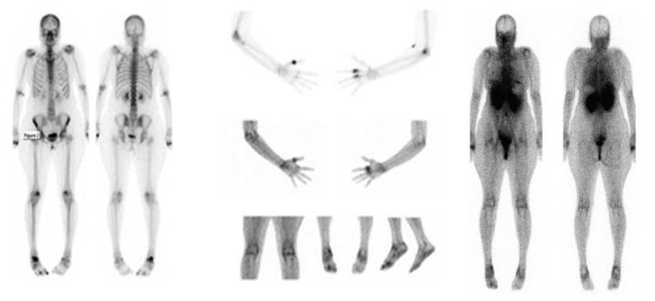
Figure 3.
Direct comparison between bone scan and glucosamine scan in a 43-year-old female with active RA. Both glucosamine scan (panel B) and bone scan (panel A) show increased tracer uptake in areas of active disease. Intensely increased bone uptake is noted in the left first MCP joint, right second and third MCP joints, proximal right radiohumeral joint, and right talonavicular joint, and is in keeping with highly active arthropathy. Less active arthropathy was seen in several left MCP joints, the left wrist, left elbow, and the patellofemoral joints. Only minimally increased uptake was noted at the shoulders. In addition, there was increased uptake involving the posterosuperior right calcaneus, which was consistent with an enthesopathy. Less active changes of enthesopathy were noted around the right cuboid, the trochanters, the proximal left radius and left navicular medially.

