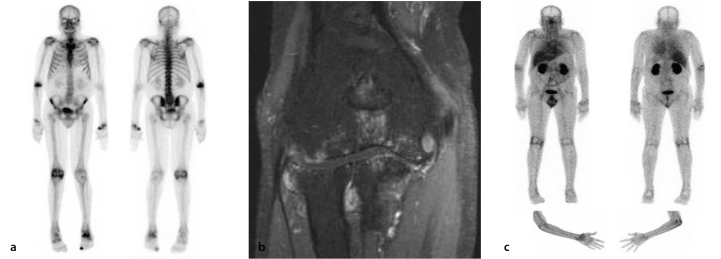
Figure 4.
Comparison between bone scan and glucosamine scan in a male RA patient with long-standing RA (15 years of disease) affecting primarily the right elbow. On the bone scan (panel A), there is increased tracer uptake (osteoblastic activity) in several joints, including the right elbow, the left wrist, and the right knee. There is a corresponding increase in 99mTc-glucosamine uptake seen in these joints on the glucosamine scans (panel C), which is consistent with active RA. Conversely, there was increased bone tracer uptake in the left ankle but minimal 99mTc-glucosamine uptake, which suggests a predominantly bone-related abnormality, rather than active RA, in the left ankle. T1 coronal fat saturation with contrast MRI (panel B) of the patient’s elbow revealed marked synovitis, effusion, and marginal erosive change with full-thickness chondral loss involving all compartments of the elbow joint, which is consistent with a long-standing erosive arthropathy with secondary osteoarthritis. Osteoarthritic changes are primarily seen in the medial compartment.

