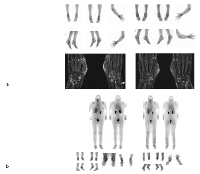
Figure 5.
Glucosamine imaging pre- and post-anti-TNF therapy. Evaluation of anti-TNF therapy on glucosamine scans. Panel A shows the glucosamine and MRI scans of a 37-year-old female with RA. Scans were carried out before commencing treatment with biologics and 8 months after treatment. The patient had clinically improved and this was reflected in the glucosamine scan, which demonstrated reduced uptake in the affected areas. Initial T1 coronal fat saturation with contrast MRI revealed marked effusions, synovitis, bony edema, erosive changes, and degenerative cyst formation involving the radiocarpal, ulnarcarpal, intercarpal, and second through fifth carpometacarpal (CMC) joints bilaterally. There was mild involvement of the first CMC and first MCP joints. Marked synovitis and effusions involving several MCP joints were most conspicuous within the second MCP and fourth proximal interphalangeal joints. Mild changes in flexor tenosynovitis were noted. Repeat T1 coronal fat saturation with contrast MRI at 8 months later revealed active erosive arthropathy, with areas of enhancement centered primarily in the carpus at the wrist and to a lesser extent in the metacarpals. The amount of edema was reduced when compared with the previous scan. Panel B of Figure 5 shows the glucosamine images of a patient with long-standing RA that had anti-TNF therapy ceased because of an infected (L) mid-foot with draining sinus. Following a period of antibiotic therapy and observation, the arthritis was exacerbated (before), requiring the recommencement of anti-TNF therapy. One year later, after commencing treatment, the patient was significantly better with no evidence of infection and 99mTc-glucosamine was reduced, particularly in the left ankle and wrists.

