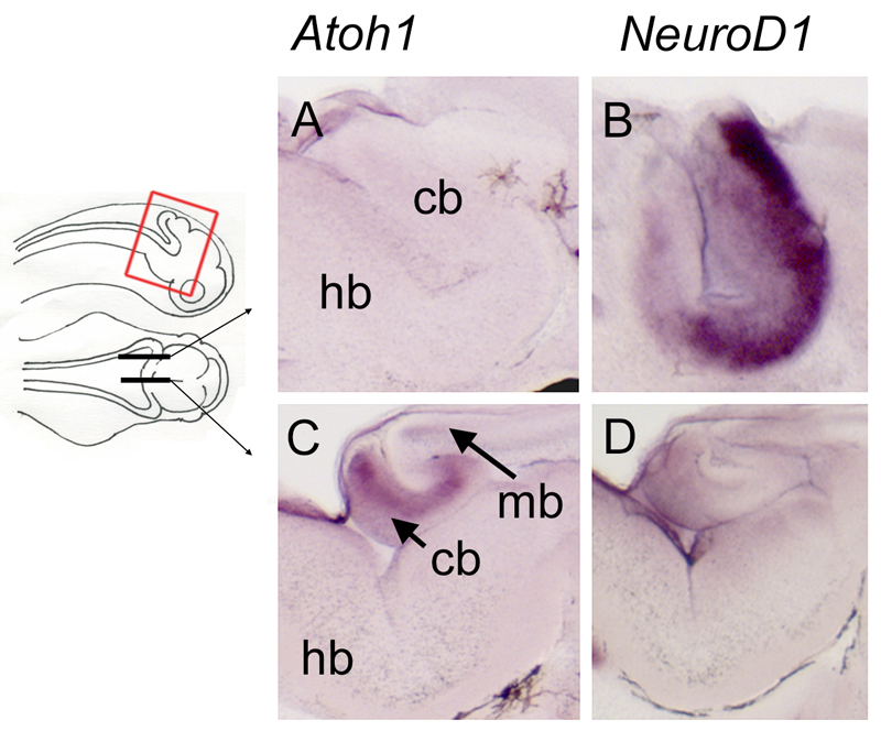
Figure 4. Complementary expression of Atoh1 and NeuroD1 in the medial versus lateral rhombic lip at stage 42.
Sections through the rhombic lip of embryos after in situ hybridisation for Atoh1 and NeuroD1 in parasagittal (A,B) and sagittal (C,D) planes, as indicated on the schematic. In the lateral rhombic lip, Atoh1-positive progenitors are absent (A) but NeuroD1 is strongly expressed (B), consistent with previously reported distributions of granule neurons (Nieuwenhuys 1967). In the medial rhombic lip, i.e., the presumptive valvulus, Atoh1 is strongly expressed (C), while NeuroD1 is reduced (D). Abbreviations: cb, cerebellum; hb, hindbrain; mb, midbrain.

