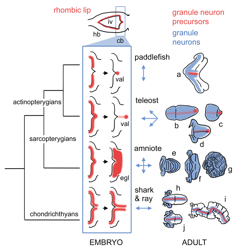
Figure 6. Hypothetical phylogeny of granule neuron proliferative zones in the cerebellum.
Hindbrain development in all vertebrate embryos can be characterised by a phase (top) in which an expanded fourth ventricle roof plate (iv) is bordered by an Atoh1-positive rhombic lip (red) in both the prospective cerebellum (cb) and the rest of the hindbrain (hb). Only the “upper” or cerebellar rhombic lip gives rise to granule cells in taxon-specific ways. We hypothesise that variation in the development of these proliferative zones (blue boxed schematics) constrains the geometry of the expansion of the granule cell layer (blue arrows, right) to produce mature granule cell distributions (blue) in adult, taxon-specific cerebellar morphologies (far right). In actinopterygians, granule cell proliferation becomes confined to a stem cell niche, the valvular primordium (val), which lies at the rostral pole of the cerebellum in teleosts (Kaslin et al. 2009) and borders the roof plate in the paddlefish. In amniotes, granule cell precursors migrate into a transient superficial external germinal layer (egl) and no precursors are retained in the adult cerebellum. In chondrichthyans, granule neuron progenitors are confined to the rhombic lip and on either side of the cerebellar midline (Chaplin, Tendeng, and Wingate 2010). Figures show hypothetical cell distributions in the cerebellar outlines (not shown to the same scale) based on model species for (a) paddlefish (a chondrostean ray-finned fish); (b-d) teleosts: b, remora (a perciform teleost), c, zebrafish (a cypriniform teleost), d, catfish (a siluriform teleost); (e-g) amniotes: e, bird, f, bat, g, giraffe; (h-j) chondrichthyans: h, dogfish (a shark), i, skate, j, stingray.
Abbreviations: cb, cerebellum; egl, external germinal layer; hb, hindbrain; iv, fourth ventricle; val, valvular primordium.

