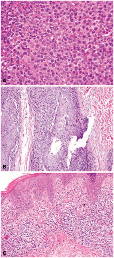
Fig. 1.

(A) H&E stain. Diffuse dermal infiltrate comprised of 100% large cells (>4× the size of a normal lymphocyte). (B) H&E stain. Hair follicles with folliculotropic infiltrate of small and large lymphocytes, at least 25% of which show large cell transformation, associated with intra-epithelial mucin deposition (follicular mucin). (C) H&E stain. Bandlike (lichenoid) infiltrate of small and large atypical lymphocytes associated with fibrotic, eosinophilic collagen bundles characteristic of chronic disease.
