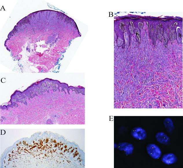
Figure 3.
Partly pigmented compound Spitz nevus with a TPM3-ALK fusion from the thigh of an 11-year-old girl (case 3). A, Wedge-shaped silhouette of a compound spindle cell melanocytic proliferation with epidermal hyperplasia (hematoxylin and eosin-stained section). B, The junctional component shows features of a pigmented spindle cell nevus. C, Deeper section of the lesion, which was adjacent to the section used for immunohistochemistry. The junctional melanocytic proliferation shows a predominant nested pattern and is pigmented. The intradermal melanocytes are amelanotic and display a plexiform growth pattern. D, The tumor cells are positive for ALK in immunohistochemistry. E, FISH confirms the ALK rearrangement.

