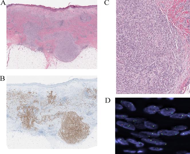
Figure 6.
Compound Spitz tumor with a DCTN1-ALK fusion from the thigh of a 9-year-old girl (case 13). A, Plaque-like intradermal melanocytic proliferation associated with a superficial dermal biopsy-related scar. The lesion shows a plexiform growth of amelanotic spindle cells with bulbous nodular growth into the superficial subcutis (hematoxylin and eosin-stained section). B, The bulbous nodule is composed of a dense proliferation of fusiform melanocytes. C, The melanocytes are immunoreactive for ALK. D, FISH confirms the ALK rearrangement.

